CN
Home > Products > Hyperspectral Imaging Camera > GaiaField-Pro Hyperspectral Imaging Camera

GaiaField-Pro

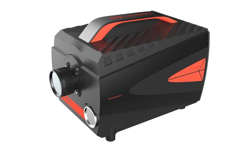
GaiaField-Pro Hyperspectral Imaging Camera

GaiaField-Pro (GFP) camera provide extremely high resolution images by scanning and taking multiple images to construct a hyperspectral datacube. Hyperspectral imaging devices (GFP) for spatial (slit) scanning obtain slit spectra by projecting a strip of the scene onto a slit and dispersing the slit image with a prism or a grating.
 
 
Inquiry
Download
Details
 
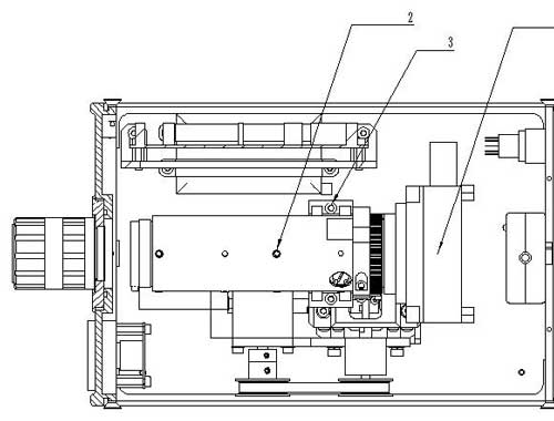
GaiaField-Pro (GFP) camera provide extremely high resolution images by scanning and taking multiple images to construct a hyperspectral datacube. Hyperspectral imaging devices (GFP) for spatial (slit) scanning obtain slit spectra by projecting a strip of the scene onto a slit and dispersing the slit image with a prism or a grating. In spatial (slit) scanning, each two-dimensional (2-D) sensor output represents a full slit spectrum (x,λ). The NUC integrated into the system to control system operation ,data acquisition, and analyze, so the spatial dimension is collected through the scanning motor movement . The line-scan systems are particularly common in remote sensing, so it is sensible to use mobile platforms. The system can keep working by internal battery about 4 hours and user can use ipad or phone to connect the camera by internal Wifi.

GaiaField Pro System Function:
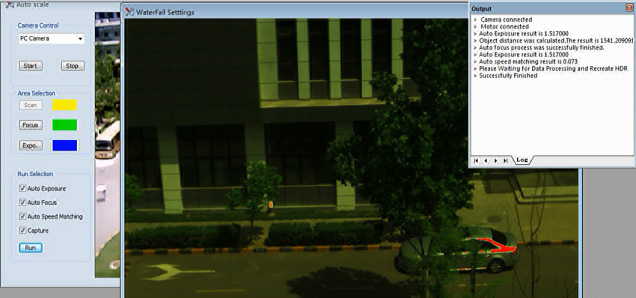
 Auto exposure time;
 Auto focusing;
 Auto match scanning speed;
 Auxiliary camera (user can observe you want );
 Data analysis (reflectance /radiometric/uniformity/lens/area calibration)
 Spectra /imaging view and export;
 Spectral Angle matching
 NDVI(advance custom)
 PCA
 Supervisedclassification
 Support lens change
 Standard type data (support envi/envice/matlab)
 Support Ipad or phone control
 
GaiaField Pro System Parameters

Instrument model

GaiaField-Pro-V10

GaiaField-Pro-V10E

GaiaField-Pro-N17E-N3

GaiaField-Pro-N17E-HR

Spectral Region(nm)

400-1000

400-1000

900-1700

900-1700

Spectral resolution(nm)

3±0.5(with 30μm slit)

2.8±0.2 (with 30μm slit)

4(with 30μm slit)

4(with 30μm slit)

Slit Length(mm)

9.6

14.2

14.2

14.2

Slit Width(μm)

30

Numerical Aperture

F/2.8

F/2.4

F/2.0

F/2.0

Light Transmission

>50%

Spectral Bands

720(2X Binning)

720(2X Binning)

100-200

200-300

Lens

Model

HSIA-OLE23

HSIA-OLES30

Focal Distance(mm)

23

30

Work Distance(mm)

100-∞

Field Angle (°)

21.5 (CCD 2/3″)

-

-

CCD Pixels

1936×1456

320x256

640×512

Pixel Size(μm)

4.54×4.54

30mm×30mm

20umx20um

Digital OutPut(bits)

8 & 12

12

14

Dynamic Range(dB)

>66

-

-

Frame Number(fps)

-

100

Exposure Time Range(ms)

0.0035-1200000

5us-1s

1us-40ms in high gain mode

Gain Mode

-

High/Low

Signal Connector

-

Ethernet port/USB

USB2.0

Lens Connector

C-Mount

Power

DC 16.8V

+12V(-0%/+5%)

12VDC

Power Consumption(W)

45

36

60

Storage Temperature(℃)

-20~+50

-45~+85

Running Temperature (℃)

+5~+40

-40~+70


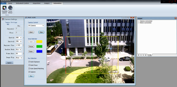
GaiaField Pro System Software:
 
 Camera settings;
 Motor settings;
 Control scanning motor;
 Control focusing motor;
 Data collect;
 Data calibration/ analysis and view;
 Saving path settings;
 Output inf and so on;
 
Fig:Specview
GaiaField Pro System Apply:
There are many applications which can take advantage of GFP hyperspectral imaging.
 Atmosphere: water vapor, cloud properties, aerosols;
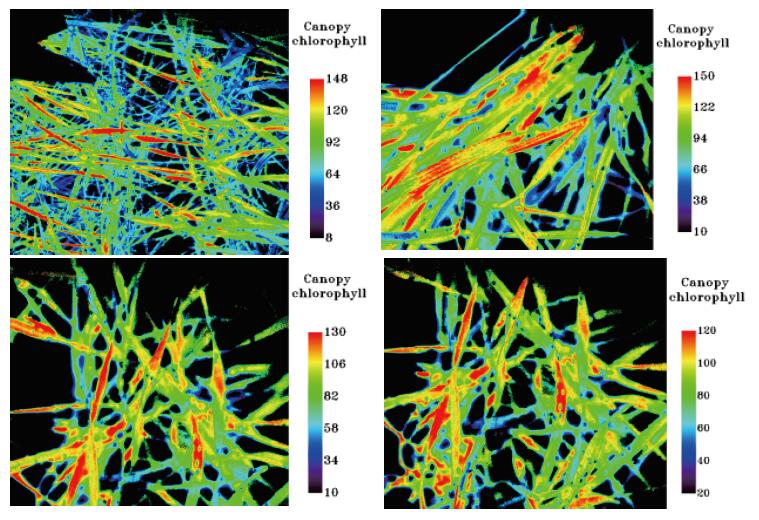
 Ecology: chlorophyll, leaf water, cellulose, pigmemts, lignin;
 Geology: mineral and soil types;
 Coastal Waters: chlorophyll,dissolved organic materials, suspended sediments;
 Snow/Ice: snow cover fraction, grainsize, melting;
 Biomass Burning: subpixel temperatures, smoke;
 Commercial: mineral exploration, agriculture and forest production
 
GaiaField Pro System Apply:
Agriculture:

 
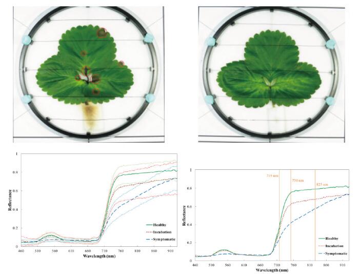
Although the cost of acquiring hyperspectral images is typically high, for specific crops and in specific climates, hyperspectral remote sensing use is increasing for monitoring the development and health of crops.Work is under way to use imaging spectrometers to detect grape variety and develop an early warning system for disease outbreaks. Furthermore, work is underway to use hyperspectral data to detect the chemical composition of plants, which can be used to detect the nutrient and water status of wheat in irrigated systems.

Medicine Care:
Researchers are working with Photon etc. and Optina Diagnostics to test the use of hyperspectral photography in the diagnosis of retinopathy and macular edema before damage to the eye occurs. The metabolic hyperspectral camera will detect a drop in oxygen consumption in the retina, which indicates potential disease. An ophthalmologist will then be able to treat the retina with injections to prevent any potential damage.
 

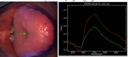
Fig .Characteristic spectrum of coating on the tongue of patients with kidney disease

Fig .Characteristic spectrum of coating on the tongue of patients with normal

Fig .Characteristic spectrum of coating on the tongue of patients with liver disease
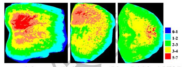
Fig .Characteristic imaging of kidney/ liver /normal tongue

Food Processing:

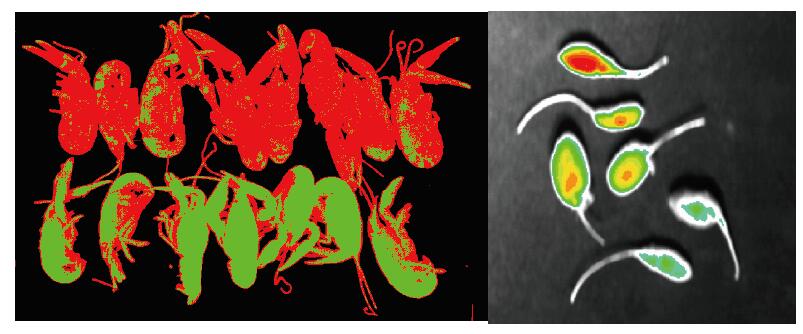
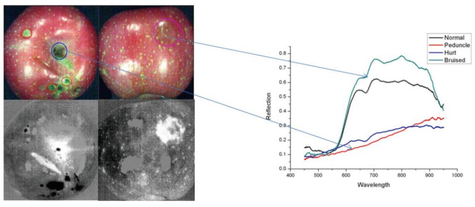
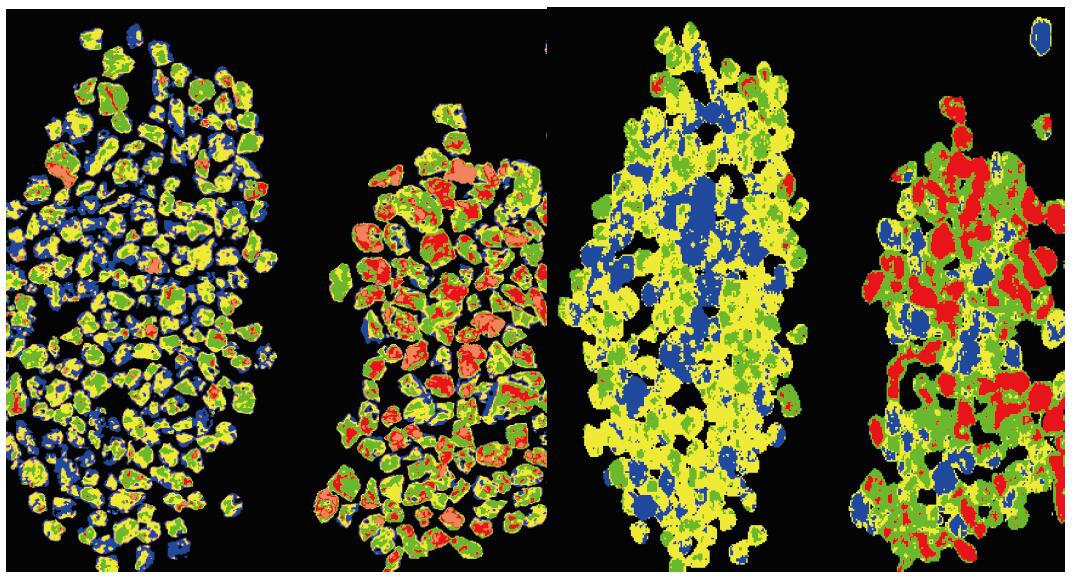
In the food processing industry, hyperspectral imaging, combined with intelligent software, enables digital sorters (also called optical sorters) to identify and remove defects and foreign material (FM) that are invisible to traditional camera and laser sorters. By improving the accuracy of defect and FM removal, the food processor’s objective is to enhance product quality and increase yields. 
Fig :(left :lobster right:lycium barbarum)
Fig: Apple Quality
Fig: Steamed Buns
Mineralogy:
 
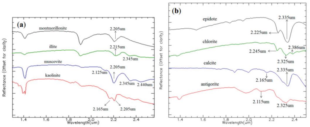
Geological samples, such as drill cores, can be rapidly mapped for nearly all minerals of commercial interest with hyperspectral imaging.

Micro imaging:
 
Combined with blood ce l l s morpho l o g i c a l characteristics and the existing method of blood cell analysis, researchers research the data of the normal blood / leukemia blood of hyperspectral imaging.The characteristics of red blood cells, lymphocytes,leukemia cells were analyzed by microscopic hyperspectral imaging, and the implementation of the diseased cells segmentation and morphological parameter quantitative calculation.


Fig: Vis-Nri Microscopic hyperspectral imaging and spectral of bacillus cell