APPLICATION
Introduction:
Endoscopy technology has been widely used in the field of pre-medical diagnosis. With the development of endoscopy technology, the interaction between light and biological tissues has play a role in early diagnosis and disease prevention. Various optical methods (such as reflection, absorption, fluorescence, scattering, etc.) have been used.
Raman spectroscopy is one of the common optical detecting methods.
1. Sample preparation is not required;
2. It is suitable for measuring living tissues with large water content;
3. Non-contact detection with biological tissues.
So, Raman Endoscopy has got many attentions and been widely used in vivo tissue diagnosis.
Raman Endoscopy technology mainly includes:
1.light source. Generally, a low-power near-infrared light source is used as the exciting light of biological tissue Raman scattering. Compared with 532nm or ultraviolet light source, it has better penetration performance, while it does less damage to biological tissues and produces less autofluorescence. At present, the wavelength of common used laser are 785nm or 830nm.
2. Endoscope probe. Exciting laser can pass through the fiber inside of the probe and generate Raman signal at the surface of tissue. It also can collect the signal, and transfer it to the optical analysis system. So, the efficiency of collecting signal, Numerical number are very important!
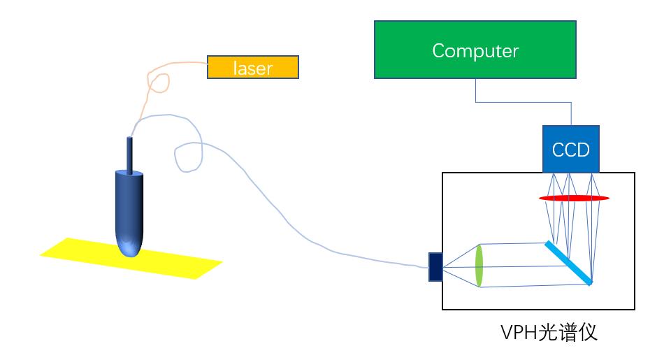
3. Optical analysis system. After the biological tissue is excited and collected by the endoscope probe, it enters the spectral analysis system through the optical fiber. Because the signal is monitored in real time, the spectral analysis system is generally based on Spectrometer and CCD (Charged Coupled Device). C-T structure and VPH (volume phase holographic) are two common configurations of spectrometer.
With the optimization and improvement of VPH gratings, it has been widely used in many Raman Endoscopy system. Compared with C-T spectrometer, it has better ability to collecting weak signals, especially the Raman signal of biological tissue. So, it can get better signal-to-noise ratio with the same optical detector. In addition, with the silicon-based CCD and 785nm or 830nm laser, the Raman signal of biological tissue can be easily measured in a wide range, not only the Fingerprint area (200-2000cm-1), but the High wavenumber range (High Wavenumber) area (2600-3500cm-1) can also be easily covered as well.
Now, there are many VPH spectrometers used in the market, and scientists have achieved good measurement results. They even can get the signals of Fingerprint and High wavenumber at the same time!
Fig.1: Schematic diagram of an endoscopic system based on a VPH spectrometer
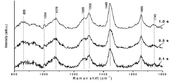
Fig.2: Comparison of in vivo Raman spectra of buccal mucosa acquired from a healthy volunteer
Under different Raman acquisition times(t=0.1, 0.5, and1.0 s)
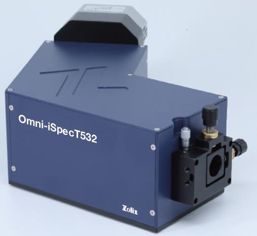
Zolix Instruments Co.Ltd. has launched a VPH spectrometer based on volume phase holographic grating !
Technical Features:
1.High Luminous Flux
F/#number: F/1.8
Perfect fiber coupling capability: 100% collection of optical signals with Fiber,NA=0.22.
2.High Light Collection Efficiency
VPH grating ensures high diffraction efficiency, and the AR-coated lens ensures the maximum light transmission efficiency in the visible or near-infrared.
3.Low Stray Light
Extremely low stray light due to smooth diffraction efficiency curve of VPH grating
Fig. 3: Diffraction Efficiency@785 Grating
4.Perfect Spectral Imaging Quality
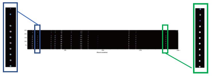
Compared with the traditional C-T mode spectrometer, the excellent optical aberration correction is achieved. We can obtain better spatial resolution and spectral resolution, and also ensure paraxial Minimal crosstalk and Raman offset for multi-channel acquisition.
Fig. 4: Neon Spectrum in Imaging Mode
20-core fiber bundle / 100μm core diameter
5.Compact and Robust design
All components are pre-adjusted as a whole module, the optical path is stable, and will not be affected by collision during transportation.
6.High Spectral Resolution
Fig. 5: Resolution Test
Application:
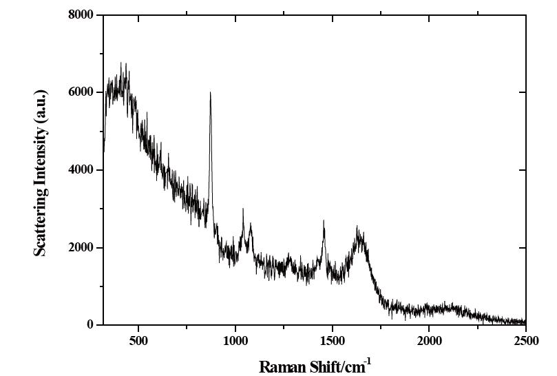
1.Ethanol solvent Raman spectroscopy (Omni-iSpecT785A1 system with high-throughput probe)
Fig. 5: Ethanol Raman spectrum, 0.25% concentration, 5s exposure time, 300mW laser
2.Human skin Raman spectroscopy (Omni-iSpecT785A1 system, high-throughput probe)
Fig. 6: Raman spectrum of skin, 20s exposure time, 20mW laser
Reference:
[1] Zhi wei Huang, Seng Khoon Teh, Wei Zheng, Opt. Lett. 34, 785 (2009).
[2] Mads Sylvest Bergholt, Wei Zheng, Zhiwei Huang, Journal of Biomedical Optics,18, 030502 (2013).
[3] Kan Lin, Wei Zheng, Chwee Ming Lim, Zhiwei Huang, Theranostics,7, 3517 (2017).